Mouse monoclonal CaMK II Antibody
CaMKII is an important member of the calcium/calmodulin-activated protein kinase family, functioning in neural synaptic stimulation and T-cell receptor signaling. CaMKII is expressed in many different tissues but is specifically found in the neurons of the forebrain and its mRNA is found within the dendrites and the soma of the neuron. The CaMKII that is found in the neurons consist of two subunits of 52 (termed alpha genes) and 60 kDa (beta genes). CaMKII has catalytic and regulatory domains, as well as an ATP-binding domain, and a consensus phosphorylation site. The binding of Ca2+/calmodulin to its regulatory domain releases its auto inhibitory effect and activates the kinase. This kinase activation results in autophosphorylation at threonine 286. The threonine phosphorylation state of CaMKII can be regulated through PP1/PKA. Whereas PP1 (protein phosphatase 1) dephosphorylates phospho-CaMKII at Thr286, PKA (protein kinase A) prevents this dephosphorylation. Autophosphorylation also enables CaMKII to attain an enhanced affinity for NMDA receptors in postsynaptic densities.

| 规格 | 价格 | 库存 |
|---|---|---|
| 100ul | 4330.00 | 15个工作日 |
基本信息
|
名称:小鼠单克隆CAMK II 抗体 描述:CaMKII is an important member of the calcium/calmodulin-activated protein kinase family, functioning in neural synaptic stimulation and T-cell receptor signaling. CaMKII is expressed in many different tissues but is specifically found in the neurons of the forebrain and its mRNA is found within the dendrites and the soma of the neuron. The CaMKII that is found in the neurons consist of two subunits of 52 (termed alpha genes) and 60 kDa (beta genes). CaMKII has catalytic and regulatory domains, as well as an ATP-binding domain, and a consensus phosphorylation site. The binding of Ca2+/calmodulin to its regulatory domain releases its auto inhibitory effect and activates the kinase. This kinase activation results in autophosphorylation at threonine 286. The threonine phosphorylation state of CaMKII can be regulated through PP1/PKA. Whereas PP1 (protein phosphatase 1) dephosphorylates phospho-CaMKII at Thr286, PKA (protein kinase A) prevents this dephosphorylation. Autophosphorylation also enables CaMKII to attain an enhanced affinity for NMDA receptors in postsynaptic densities. 克隆:单克隆 22B1 同种型:IgG1 宿主:Mouse 别名:CSAID Binding protein 1 Antibody, CSBP1 Antibody, CSBP2 Antibody, EXIP Antibody, MAP kinase MXI2 Antibody, MAPkinase p38alpha Antibody, MAPK14 Antibody, p38 ALPHA Antibody, p38 MAP kinase Antibody, p38 mitogen activated protein kinase Antibody, RK Antibody, SAPK 2A Antibody, Stress activated protein kinase 2A Antibody 分子量:73 kDa 种属反应性:Human, Mouse, Rat 经测试应用:WB,IHC-P/IHC-Fr,IF/ICC,IP,ELISA 浓度:1mg/ml 纯化方式:Protein G Purified. 抗原:Synthetic peptide to Rat CaMKII. 浓度:WB:1:500-1000 IHC-P/IHC-Fr:1:50-200 IF/ICC:1:500-1000 存储液:PBS pH7.4, 50% glycerol, 0.09% sodium azide. 储存温度:-20℃,避免污染
Western Blot analysis of Mouse Ventricle lysates showing detection of CaMKII protein using Mouse monoclonal CaMK II Antibody at 1:1000. Analysis of CaMKII and NFAT phosphorylation in ventricles of 14 day old mice over-expressing CaMK.
Immunocytochemistry/Immunofluorescence analysis using Mouse monoclonal CaMK II Antibody. Tissue: dissociated hippocampal neurons. Species: Rat. Fixation: Cold 4% paraformaldehyde/0.2% glutaraldehyde in 0.1M sodium phosphate buffer. Primary Antibody: Mouse monoclonal CaMK II Antibody at 1:1000 for 12 hours at 4°C. Secondary Antibody: FITC Goat Anti-Mouse IgG (green) at 1:50 for 30 minutes at RT. Magnification: 10X.
Immunohistochemistry analysis using Mouse monoclonal CaMK II Antibody. Tissue: backskin. Species: Mouse. Fixation: Bouin’s Fixative and paraffin-embedded. Primary Antibody: Mouse monoclonal CaMK II Antibody at 1:100 for 1 hour at RT. Secondary Antibody: FITC Goat Anti-Mouse (green) at 1:50 for 1 hour at RT. Localization: Muscle, hair follicle, epidermis. Backskin obtained from transgenic mice.
Immunohistochemistry analysis using Mouse monoclonal CaMK II Antibody. Tissue: colon carcinoma. Species: Human. Fixation: Formalin. Primary Antibody: Mouse monoclonal CaMK II Antibody at 1:5000 for 12 hours at 4°C. Secondary Antibody: Biotin Goat Anti-Mouse at 1:2000 for 1 hour at RT. Counterstain: Mayer Hematoxylin (purple/blue) nuclear stain at 200 µl for 2 minutes at RT. Magnification: 40x. |
|
| 存储温度 | -20℃ |
相关说明文档
储备液配置
相关产品推荐
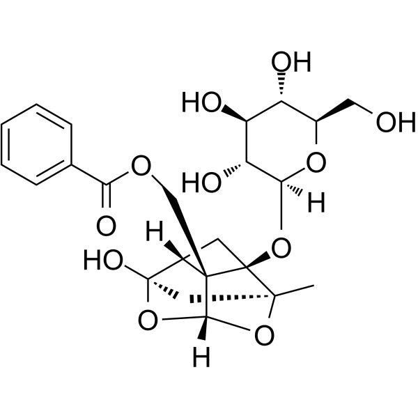
Peoniflorin(芍药苷)是来自于芍药的根部,在中医理论上是有止痛属性,抗心肌缺血,镇痛,抗炎,耐缺氧等作用。
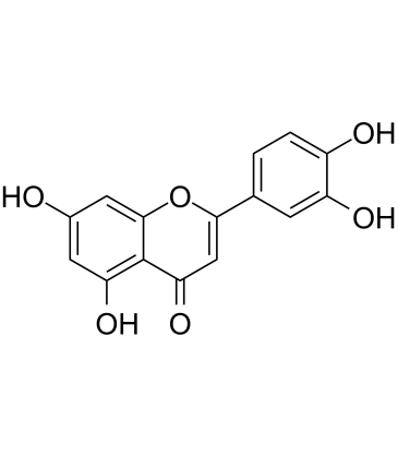
Luteolin is a non-selective phisphodiesterase PDE inhibitor
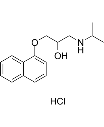
Propranolol hydrochloride 是一种非选择性的肾上腺素受体拮抗剂,Propranolol hydr
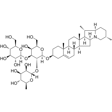
α-solanine 是马铃薯中的一种生物活性成分,Alpha-Solanine可改变脂肪和肠中的抗氧化酶活性,MDA和


